Topic 1.4 Notes – The Brain
What the Brain Does
The brain coordinates behavior and mental processes. That includes obvious actions like moving your hand and less visible things like memory, emotion, language, and judgment.
Two ideas have to stay together:
- Localization of function means some brain regions are especially linked to certain jobs.
- Example: the occipital lobe is strongly tied to vision.
- Integration means most behavior uses networks, not one isolated spot.
- Example: speaking involves language areas, motor control, hearing, and attention.
A common AP move is giving you a symptom and asking for the structure. Your job is to match the main loss or change to the brain area most associated with it.
This brain diagram is a good quick reference for the big regions you will keep seeing, especially the lobes, cerebellum, and brain stem.
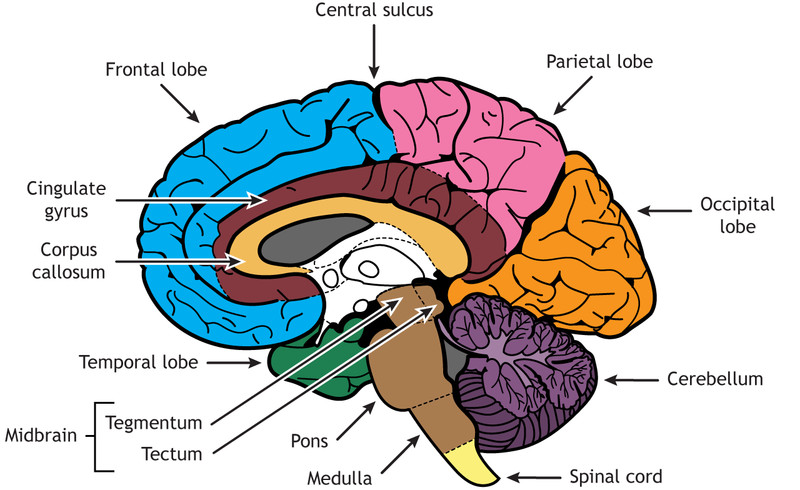
Major brain regions
Major Brain Structures and What They Control
Brain stem, reticular formation, reward system, and cerebellum
- Brain stem handles basic survival functions.
- Medulla regulates breathing and heart rate.
- Reticular formation or reticular activating system helps with alertness, arousal, attention, eye movement, and some movement. Major damage can reduce consciousness.
- Reward center means a dopamine-linked network involved in reinforcement, motivation, some learning, cognition, and emotion. It is a network, not one tiny “pleasure spot.”
- Cerebellum helps balance, coordination, smooth voluntary movement, and some procedural learning like learned skills.
Limbic system
This group is big for emotion, memory, motivation, and body regulation.
- Thalamus relays most sensory information to the right cortical area. Smell is the main exception.
- Hypothalamus maintains homeostasis such as hunger, thirst, and body temperature. It also links the nervous and endocrine systems.
- Pituitary gland is the “master gland” that releases hormones under hypothalamic control.
- Hippocampus forms new explicit memories for facts and events.
- Amygdala is tied to fear, threat detection, aggression, and emotional significance.
Cerebral cortex and the four lobes
The cerebral cortex is the outer layer for complex processing and has two hemispheres.
- Occipital lobes process vision.
- Temporal lobes process hearing and language comprehension.
- Parietal lobes help integrate information and contain the somatosensory cortex, which maps body touch sensations. This map is contralateral, so the left hemisphere processes the right side of the body.
- Frontal lobes handle language, planning, judgment, higher-order thinking, and voluntary movement.
- Prefrontal cortex supports executive functions like planning, decision-making, and impulse control.
- Motor cortex controls skeletal movement and also works contralaterally.
The labeled cortical map below connects these lobe functions to specific areas you should recognize, especially the motor cortex, somatosensory cortex, visual cortex, auditory cortex, and the language areas Broca’s and Wernicke’s.
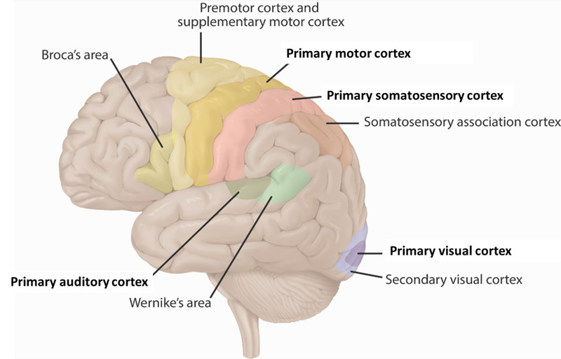
Hemispheres, Language, and Split-Brain Findings
The corpus callosum is the fiber bundle connecting the hemispheres. It lets the two sides share information.
Lateralization means some functions are more associated with one hemisphere:
- Left hemisphere is usually language dominant.
- Right hemisphere is often stronger in spatial and perceptual tasks.
Do not fall for the myth that people are simply “left-brained” or “right-brained.”
Language areas are usually on the left:
- Broca’s area in the left frontal lobe helps speech production.
- Wernicke’s area in the left temporal lobe helps language comprehension.
- Aphasia means language impairment from brain damage.
- Broca’s patient “Tan” helped show localization of speech production.
Split-brain research showed this step by step. In the illustration, focus on the idea that information shown to one visual field is processed by the opposite hemisphere.
- The corpus callosum was severed to reduce severe epilepsy.
- Sperry and Gazzaniga studied these patients.
- Right visual field → left hemisphere so the person can usually name the object.
- Left visual field → right hemisphere so a split-brain patient may not verbally name it.
- The left hand can still point to it because the right hemisphere processed it.
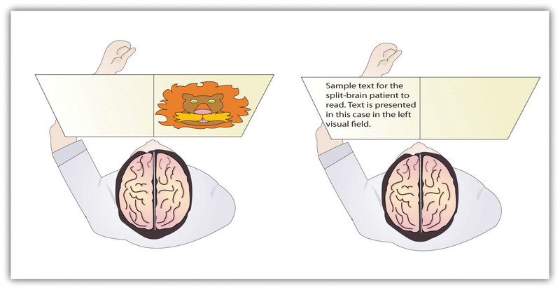
Split-brain visual field testing
Damage, Plasticity, and Classic Case Studies
Neuroplasticity is the brain’s ability to rewire or change connections. It is strongest early in life but continues in adulthood. It can support recovery after damage, though recovery is often partial.
Classic cases:
- Phineas Gage had frontal lobe damage linked to personality, judgment, and impulse-control changes.
- Henry Molaison (H.M.) had hippocampal damage and could not form new explicit memories, though some procedural learning remained.
Fast matches you should know:
- breathing or heart rate → medulla
- alertness loss → reticular formation
- poor balance → cerebellum
- vision problems → occipital lobe
- comprehension problems → temporal lobe or Wernicke’s area
- touch problems → parietal lobe or somatosensory cortex
- movement problems on opposite side → motor cortex
- poor planning or impulse control → prefrontal cortex
- no new explicit memories → hippocampus
- abnormal fear or aggression → amygdala
- homeostasis problems → hypothalamus
How Psychologists Study the Brain
Different methods answer different questions.
| Method | Best at | Limitation |
|---|---|---|
| EEG | timing of electrical activity | weak location precision |
| fMRI | where activity is, using blood-oxygen changes | weaker timing, indirect measure |
| Lesions/surgery | showing what happens when tissue is damaged or removed | damage may be messy unless tightly controlled |
| Case studies | rich detail from rare patients | low generalizability |
The best conclusion uses converging evidence. If imaging, lesions, and case studies all point to the same function, the claim is much stronger.
Key Takeaways
Localization of Function
The principle that particular brain regions are especially associated with particular functions, although behavior usually depends on integrated networks
Brain Stem
The structure connecting the spinal cord to the rest of the brain that generally controls basic life-sustaining functions
Medulla
The part of the brain stem that automatically regulates breathing and heart rate
Reticular Activating System (Reticular Formation)
A brain-stem network that regulates alertness, arousal, attention, eye movement, and some voluntary movement
Reward Center
Interconnected brain regions whose activity makes experiences rewarding, reinforces associated behavior, and contributes to motivation, learning, cognition, and emotion
Cerebellum
The lower-rear brain structure that coordinates smooth voluntary movement, balance, and some procedural learning
Cerebral Cortex
The brain’s folded outer layer, divided into two hemispheres, that supports sensory processing, voluntary movement, language, reasoning, and planning
Occipital Lobes
The lobes at the rear of the cerebral cortex that primarily process visual information
Temporal Lobes
The lobes on the sides of the cerebral cortex that primarily support auditory and linguistic processing
Parietal Lobes
The lobes near the upper rear of the cortex that organize and integrate information and contain the somatosensory cortex
Association Areas
Cortical regions that integrate and organize information rather than receiving one basic sensory signal or issuing one basic motor command
Somatosensory Cortex
A strip at the front of each parietal lobe that processes touch and other body sensations, mainly from the opposite side
Frontal Lobes
The lobes behind the forehead that support language, voluntary movement, higher-order thinking, and executive functioning
Executive Functions
Higher-order processes including planning, judgment, attention control, problem solving, impulse inhibition, and goal-directed regulation
Prefrontal Cortex
The front portion of the frontal lobes that is especially important for planning, judgment, impulse control, and behavioral regulation
Motor Cortex
A strip at the rear of each frontal lobe that controls most skeletal movement, mainly on the opposite side of the body
Limbic System
An interconnected system including the thalamus, hypothalamus, pituitary gland, hippocampus, and amygdala that supports memory, emotion, motivation, and internal regulation
Thalamus
The major relay station that directs incoming sensory information, except smell, to appropriate cortical areas
Hypothalamus
A structure that regulates hunger, thirst, body temperature, and other homeostatic processes and directs the pituitary gland
Pituitary Gland (Master Gland)
The hypothalamus-regulated gland that releases hormones influencing bodily processes and the activity of other endocrine glands
Hippocampus
A limbic structure essential for processing and consolidating new explicit memories
Amygdala
A limbic structure involved in emotional significance, emotional learning, fear, threat detection, and aggression
Corpus Callosum
The large bundle of axons that connects the cerebral hemispheres and allows them to exchange information
Lateralization (Hemispheric Specialization)
The tendency for some functions to be more strongly associated with one cerebral hemisphere than the other
Contralateral Organization
The organization in which each cerebral hemisphere primarily processes or controls information from the opposite side
Broca’s Area
A region usually in the left frontal lobe responsible for speech production; damage commonly causes slow, effortful, nonfluent speech
Wernicke’s Area
A region usually in the left temporal lobe responsible for language comprehension; damage can cause fluent but incoherent speech and poor understanding
Aphasia
An impairment of language production, comprehension, or both caused by brain damage
Split-Brain Research
Research on patients with severed corpus callosa that presents stimuli to one visual field to reveal contralateral processing and hemispheric specialization
Brain Plasticity (Neuroplasticity)
The brain’s ability to modify, strengthen, weaken, or create neural connections through development, experience, learning, or recovery from injury
Electroencephalogram (EEG)
A noninvasive scalp recording of electrical brain activity with high temporal resolution but limited spatial precision
Functional Magnetic Resonance Imaging (fMRI)
A noninvasive method that indirectly maps brain activity through blood-oxygen and blood-flow changes, with strong spatial but weaker temporal resolution
Case Study
An intensive investigation of one person or a few unusual cases that provides detail but has limited control and generalizability
Lesion
Damaged or destroyed brain tissue studied to infer a region’s function from the behavioral capacities that change or remain intact
Direct Brain Stimulation
Applying stimulation to exposed brain tissue during medically justified surgery to observe responses and map functions
Phineas Gage
A railroad foreman whose frontal-lobe injury was followed by changes in judgment, impulse control, personality, and social behavior
Henry Molaison (Patient H.M.)
A patient whose bilateral hippocampal surgery severely impaired new explicit-memory formation while leaving some older memories and procedural learning intact
Notes
Localization of Function
The principle that particular brain regions are especially associated with particular functions, although behavior usually depends on integrated networks
Brain Stem
The structure connecting the spinal cord to the rest of the brain that generally controls basic life-sustaining functions
Medulla
The part of the brain stem that automatically regulates breathing and heart rate
Reticular Activating System (Reticular Formation)
A brain-stem network that regulates alertness, arousal, attention, eye movement, and some voluntary movement
Reward Center
Interconnected brain regions whose activity makes experiences rewarding, reinforces associated behavior, and contributes to motivation, learning, cognition, and emotion
Cerebellum
The lower-rear brain structure that coordinates smooth voluntary movement, balance, and some procedural learning
Cerebral Cortex
The brain’s folded outer layer, divided into two hemispheres, that supports sensory processing, voluntary movement, language, reasoning, and planning
Occipital Lobes
The lobes at the rear of the cerebral cortex that primarily process visual information
Temporal Lobes
The lobes on the sides of the cerebral cortex that primarily support auditory and linguistic processing
Parietal Lobes
The lobes near the upper rear of the cortex that organize and integrate information and contain the somatosensory cortex
Association Areas
Cortical regions that integrate and organize information rather than receiving one basic sensory signal or issuing one basic motor command
Somatosensory Cortex
A strip at the front of each parietal lobe that processes touch and other body sensations, mainly from the opposite side
Frontal Lobes
The lobes behind the forehead that support language, voluntary movement, higher-order thinking, and executive functioning
Executive Functions
Higher-order processes including planning, judgment, attention control, problem solving, impulse inhibition, and goal-directed regulation
Prefrontal Cortex
The front portion of the frontal lobes that is especially important for planning, judgment, impulse control, and behavioral regulation
Motor Cortex
A strip at the rear of each frontal lobe that controls most skeletal movement, mainly on the opposite side of the body
Limbic System
An interconnected system including the thalamus, hypothalamus, pituitary gland, hippocampus, and amygdala that supports memory, emotion, motivation, and internal regulation
Thalamus
The major relay station that directs incoming sensory information, except smell, to appropriate cortical areas
Hypothalamus
A structure that regulates hunger, thirst, body temperature, and other homeostatic processes and directs the pituitary gland
Pituitary Gland (Master Gland)
The hypothalamus-regulated gland that releases hormones influencing bodily processes and the activity of other endocrine glands
Hippocampus
A limbic structure essential for processing and consolidating new explicit memories
Amygdala
A limbic structure involved in emotional significance, emotional learning, fear, threat detection, and aggression
Corpus Callosum
The large bundle of axons that connects the cerebral hemispheres and allows them to exchange information
Lateralization (Hemispheric Specialization)
The tendency for some functions to be more strongly associated with one cerebral hemisphere than the other
Contralateral Organization
The organization in which each cerebral hemisphere primarily processes or controls information from the opposite side
Broca’s Area
A region usually in the left frontal lobe responsible for speech production; damage commonly causes slow, effortful, nonfluent speech
Wernicke’s Area
A region usually in the left temporal lobe responsible for language comprehension; damage can cause fluent but incoherent speech and poor understanding
Aphasia
An impairment of language production, comprehension, or both caused by brain damage
Split-Brain Research
Research on patients with severed corpus callosa that presents stimuli to one visual field to reveal contralateral processing and hemispheric specialization
Brain Plasticity (Neuroplasticity)
The brain’s ability to modify, strengthen, weaken, or create neural connections through development, experience, learning, or recovery from injury
Electroencephalogram (EEG)
A noninvasive scalp recording of electrical brain activity with high temporal resolution but limited spatial precision
Functional Magnetic Resonance Imaging (fMRI)
A noninvasive method that indirectly maps brain activity through blood-oxygen and blood-flow changes, with strong spatial but weaker temporal resolution
Case Study
An intensive investigation of one person or a few unusual cases that provides detail but has limited control and generalizability
Lesion
Damaged or destroyed brain tissue studied to infer a region’s function from the behavioral capacities that change or remain intact
Direct Brain Stimulation
Applying stimulation to exposed brain tissue during medically justified surgery to observe responses and map functions
Phineas Gage
A railroad foreman whose frontal-lobe injury was followed by changes in judgment, impulse control, personality, and social behavior
Henry Molaison (Patient H.M.)
A patient whose bilateral hippocampal surgery severely impaired new explicit-memory formation while leaving some older memories and procedural learning intact
