Topic 2.4 Notes – Membrane Permeability
Selective permeability and the hydrophobic barrier
Every cell needs to keep its internal environment different from the outside. The plasma membrane makes that possible by separating the cytoplasm from the external environment.
Selective permeability means:
- Some molecules cross easily.
- Others cross slowly or not at all.
- Some require specific proteins.
The reason comes down to the membrane’s hydrophobic interior.
Inside the membrane, the fatty acid tails of phospholipids form a nonpolar core. Nonpolar substances can dissolve in this region. Polar and charged substances cannot.
So the membrane does not actively “block” most molecules. Its structure naturally creates a barrier.
Big connection:
Structure → hydrophobic core → selective permeability → homeostasis
On quizzes, you’ll often be asked to explain permeability in terms of structure, not just state what can cross.
What the plasma membrane is made of
Phospholipid bilayer
Phospholipids are amphipathic. That means they have two different regions:
- Hydrophilic (polar) phosphate head → interacts with water
- Hydrophobic (nonpolar) fatty acid tails → avoid water
In water, they form a bilayer with heads facing the extracellular and intracellular fluids and tails tucked toward each other in the center.
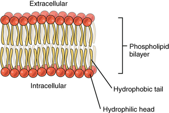
Phospholipid bilayer structure
That inward-facing tail region is the hydrophobic barrier that determines permeability.
Membrane proteins
Proteins are embedded throughout the bilayer. Their placement depends on their amino acids:
- Hydrophobic regions sit in the membrane interior.
- Hydrophilic regions face water.
Important types for permeability:
- Channel proteins
- Form hydrophilic tunnels
- Allow specific ions or polar molecules to move across
- Passive transport
- Transport proteins (carriers/pumps)
- Bind substances and change shape to move them
- Some use ATP (active transport, covered more in 2.5)
Other membrane proteins you should recognize:
- Receptor proteins → receive signals
- Adhesion proteins → attach cells together
- Cell surface markers (glycoproteins) → cell ID
For this topic, channel and transport proteins matter most because they allow hydrophilic substances to bypass the hydrophobic core.
Fluid mosaic model
The membrane is described as a fluid mosaic:
- Fluid → phospholipids and many proteins move laterally.
- Mosaic → mixture of lipids and proteins embedded together.
This flexibility allows proteins to function and the membrane to self-repair.
What can and cannot cross the membrane
Permeability depends on size, polarity, and charge.
Freely cross
Small, nonpolar molecules dissolve in the hydrophobic interior:
- O₂
- CO₂
- N₂
They cross by simple diffusion.
Cross slowly in small amounts
Small, polar, uncharged molecules:
- H₂O
- NH₃
They can slip through the bilayer, but slowly. In cells, water mainly uses aquaporin channel proteins for efficient movement.
Cannot cross without proteins
- Ions (Na⁺, Cl⁻, Ca²⁺)
- Large polar molecules (glucose)
The nonpolar hydrocarbon tails repel these substances. They require channel or transport proteins.
Here’s a clean comparison:
| Molecule Type | Polarity/Charge | Crosses Freely? | Needs Protein? |
|---|---|---|---|
| O₂, CO₂, N₂ | Small, nonpolar | Yes | No |
| H₂O, NH₃ | Small, polar, uncharged | Small amounts | Often yes |
| Na⁺, Cl⁻, glucose | Charged or large polar | No | Yes |
A common AP move is giving you a new molecule and asking you to predict movement based on polarity and charge. Always think about the hydrophobic core.
The role of the cell wall
Some organisms have a cell wall outside the plasma membrane.
Found in:
- Plants → cellulose
- Fungi → chitin
- Bacteria → peptidoglycan
- Archaea → other polymers
Animal cells do not have cell walls.
Functions:
- Structural support → maintains shape
- Protection from osmotic lysis → prevents bursting in hypotonic environments
- Additional permeability barrier → filters some substances before they reach the membrane
In plant cells, you can see the thick cell wall forming the rigid outer boundary, with the plasma membrane just inside it.
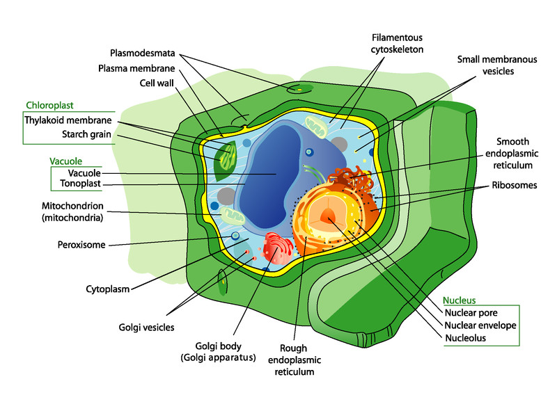
Labeled plant cell showing the cell wall outside the plasma membrane
In a hypotonic environment, water enters the cell. The plasma membrane allows water movement, but the rigid cell wall prevents overexpansion and bursting.
So the membrane controls molecular movement. The wall provides structural protection and an extra layer of filtering.
Key Takeaways
Fluid Mosaic Model
A dynamic membrane model with moving phospholipids and embedded proteins forming a flexible mosaic.
Transport Proteins and Channel Proteins
Embedded membrane proteins that move hydrophilic substances across the bilayer; channels are passive, transport proteins may actively pump.
Plasma Membrane
The cell boundary that separates internal conditions from the external environment.
Phospholipid Bilayer
A double phospholipid layer with a hydrophobic interior that causes selective permeability.
Selective Permeability
A membrane property where small nonpolar molecules cross easily but polar molecules and ions do not.
Cell Wall
A rigid outer layer that supports cells, limits some substances, and prevents osmotic lysis.
Notes
Fluid Mosaic Model
A dynamic membrane model with moving phospholipids and embedded proteins forming a flexible mosaic.
Transport Proteins and Channel Proteins
Embedded membrane proteins that move hydrophilic substances across the bilayer; channels are passive, transport proteins may actively pump.
Plasma Membrane
The cell boundary that separates internal conditions from the external environment.
Phospholipid Bilayer
A double phospholipid layer with a hydrophobic interior that causes selective permeability.
Selective Permeability
A membrane property where small nonpolar molecules cross easily but polar molecules and ions do not.
Cell Wall
A rigid outer layer that supports cells, limits some substances, and prevents osmotic lysis.
