Topic 1.7 Notes – Proteins
1. What Proteins Are
Proteins are polymers of amino acids. Each amino acid is a monomer, and when many are linked together, they form a polypeptide, which folds into a functional protein.
Peptide Bonds and Dehydration Synthesis
Amino acids are connected by covalent peptide bonds.
Here’s what actually happens chemically:
- The carboxyl group (-COOH) of one amino acid reacts with
- The amino group (-NH₂) of another
- A molecule of water (H₂O) is removed
- A peptide bond forms between them
This is a dehydration synthesis reaction.
As more amino acids join, the chain grows in a specific order. That order is determined by genetic information in DNA. The cell reads a gene, builds a polypeptide in that exact sequence, and then the chain folds.
That folding is where structure and function begin to connect.
Big idea:
Sequence → Structure → Function
Change the sequence, and you can change the structure. Change the structure, and you can change the function.
2. Amino Acids and Their R Groups
Basic Structure of an Amino Acid
All amino acids share the same core structure. The diagram below shows the general layout of a single amino acid.

General structure of an amino acid
Every amino acid has:
- A central (alpha) carbon
- A hydrogen
- An amino group (-NH₂)
- A carboxyl group (-COOH)
- A variable R group (side chain)
The R group is what makes each amino acid chemically unique. You do not need to memorize detailed molecular structures for AP, but you must understand how R groups behave.
Three Categories of R Groups
Here’s how AP wants you to think about them:
| Type | Property | Effect on Protein |
|---|---|---|
| Nonpolar (Hydrophobic) | Repel water | Cluster inside the protein; stabilize via hydrophobic interactions |
| Polar (Hydrophilic) | Interact with water | Often found on surfaces; form hydrogen bonds |
| Ionic (Charged) | Positive or negative charge | Form ionic bonds (salt bridges); affect shape and stability |
The interactions between these R groups drive folding. A single amino acid substitution can disrupt those interactions. That’s exactly what happens in sickle-cell disease, where one amino acid change alters hemoglobin’s structure.
This idea shows up constantly in FRQs. If a mutation changes an amino acid from nonpolar to charged, you’re expected to predict how that might disrupt folding or interactions.
3. The Four Levels of Protein Structure
Each level builds on the one before it.
Primary Structure
- The linear sequence of amino acids
- Held together by peptide bonds
- Directly determined by DNA
Everything else depends on this level.
Secondary Structure
Secondary structure is local folding of the backbone, not the R groups.
It forms due to hydrogen bonds between atoms in the polypeptide backbone.
Two main shapes are shown below.
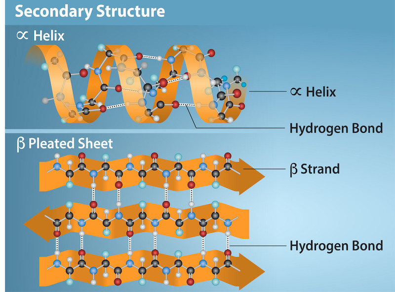
Alpha helix and beta pleated sheet secondary structures
- Alpha helix (α-helix) → spiral coil stabilized by backbone hydrogen bonds
- Beta pleated sheet (β-sheet) → folded strands held together by backbone hydrogen bonds
Students often think R groups cause this level. They do not. It’s backbone hydrogen bonding.
Tertiary Structure
This is the overall 3D shape of a single polypeptide.
It forms due to R-group interactions, including:
- Hydrogen bonds
- Ionic bonds
- Hydrophobic interactions
- Disulfide bridges (covalent bonds between cysteines)
This level largely determines function. If an FRQ describes changes in pH or temperature, you should think about disruption of hydrogen bonds or ionic interactions at this level.
Quaternary Structure
Some proteins contain multiple polypeptide subunits.
Quaternary structure results from interactions between those subunits.
- Same types of interactions as tertiary structure
- Not all proteins have this level
- Example: Hemoglobin (4 subunits)
All four levels together determine final function.
4. How Protein Structure Determines Function
Protein shape determines:
- Binding specificity
- Catalytic activity (enzymes)
- Stability
- Interactions with other molecules
Changes that can alter structure:
- Amino acid substitution
- pH changes
- Temperature increases
- Disruption of disulfide bonds
When proteins misfold, they may lose function or aggregate, leading to disease.
On the AP exam, you’ll often be given a mutation and asked to explain how it affects protein function. The scoring almost always requires you to trace the path:
DNA change → amino acid change → altered interactions → changed structure → altered function.
5. Major Functions of Proteins
Proteins do almost everything in cells:
- Enzymes → speed up chemical reactions
- Structural proteins → collagen, keratin
- Transport proteins → hemoglobin
- Signaling proteins → insulin
- Defense proteins → antibodies
- Movement proteins → actin and myosin
Different functions, same rule: specific structure allows specific interactions.
And that structure ultimately comes from the amino acid sequence encoded by DNA.
Key Takeaways
Amino Acid Structure
A central carbon bonded to an amino group, carboxyl group, hydrogen, and variable R group.
R Group Categories
Nonpolar hydrophobic, polar hydrophilic, and ionic charged side chains with different chemical behaviors.
Peptide Bond
A covalent bond linking one amino acid's carboxyl group to another's amino group.
Polypeptide Chain
A linear polymer of amino acids connected by peptide bonds.
Primary Structure
The specific linear sequence of amino acids in a polypeptide.
Secondary Structure
Local folding of the polypeptide backbone into alpha helices and beta pleated sheets by hydrogen bonding.
Tertiary Structure
The overall three-dimensional shape of one polypeptide formed by side-chain interactions.
Tertiary Structure Interactions
Hydrogen bonds, ionic interactions, hydrophobic interactions, and disulfide bridges stabilize one polypeptide's shape.
Quaternary Structure
The association of multiple polypeptide subunits into one functional protein.
Protein Functions
Common roles include catalysis, structure, transport, signaling, defense, and movement.
Disulfide Bridge
A covalent bond between sulfur-containing side chains that stabilizes a protein's folded shape.
Notes
Amino Acid Structure
A central carbon bonded to an amino group, carboxyl group, hydrogen, and variable R group.
R Group Categories
Nonpolar hydrophobic, polar hydrophilic, and ionic charged side chains with different chemical behaviors.
Peptide Bond
A covalent bond linking one amino acid's carboxyl group to another's amino group.
Polypeptide Chain
A linear polymer of amino acids connected by peptide bonds.
Primary Structure
The specific linear sequence of amino acids in a polypeptide.
Secondary Structure
Local folding of the polypeptide backbone into alpha helices and beta pleated sheets by hydrogen bonding.
Tertiary Structure
The overall three-dimensional shape of one polypeptide formed by side-chain interactions.
Tertiary Structure Interactions
Hydrogen bonds, ionic interactions, hydrophobic interactions, and disulfide bridges stabilize one polypeptide's shape.
Quaternary Structure
The association of multiple polypeptide subunits into one functional protein.
Protein Functions
Common roles include catalysis, structure, transport, signaling, defense, and movement.
Disulfide Bridge
A covalent bond between sulfur-containing side chains that stabilizes a protein's folded shape.
